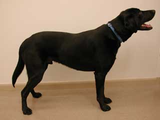
| This is a picture of a Labrador retriever that has normal neurological function. Notice that the dog is alert and responsive, does not have a head tilt, does not drift or lean to one side, and places equal weight on all limbs. |
pupillary light reflex test (plr) | menace reaction | patellar reflex test | triceps reflex | deep pain
Introduction: There are many different diseases and problems that can affect an animal’s neurological system (the brain, spinal cord, and nerves). The following information and pictures will help an owner identify when the nervous system is functioning normally and will be particularly helpful in identifying when there is a neurological problem. When a neurological problem has been identified, it is essential that the pet be taken to a veterinarian for immediate examination and proper treatment. Failure to act promptly when a problem is identified can lead to serious and permanent damage. It is also essential to use extreme caution when examining or moving an injured animal. The injured pet may try to protect itself from additional injury by biting.
To begin the examination, look at the pet from a distance. Notice how the pet is standing, walking, and holding its head. Notice any head tilt, leaning or drifting to one side, short or choppy steps, or general weakness as the pet moves. This is a picture of a normal dog, standing with all four feet, the head, and body in proper position.
|
|
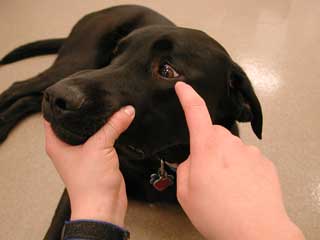
Next, observe and handle the dog’s head and mouth areas. It is sometimes helpful to watch the dog eat if there is a problem with the dog prehending (obtaining) food, chewing, or swallowing. While examining the head, function of the cranial nerves can be evaluated. There are 12 cranial nerves that are numbered I through XII. These cranial nerves are responsible for a variety of special senses and functions, including eye movement and position (III, IV, and VI), feeling and motor function to the face and head (V and VII), balance and hearing (VIII), taste and feeling to the back of the throat (IX), swallowing and some control over digestion and heart function (X), some muscular movement to shoulders and head (XI), and movement of the tongue (XII). Cranial nerve I aids in the sense of smell and cranial nerve II helps with sight/vision.
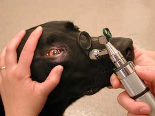
The following photos show how to evaluate some of the cranial nerves. Only a few of the more basic tests will be outlined here. What cranial nerve is being tested and what a normal response would be have been included with each picture.
 |
|
 |
|
General eye examination: Both pupils should be the same size and should not be too dilated or constricted. The eyes should be able to follow basic movements and distractions. An involuntary, rhythmic movement of the eye called nystagmus can also be produced in a normal eye. This nystagmus can be seen by gently turning the animals head from side to side in a horizontal fashion. A normal nystagmus response is seen when the eyes move slowly away from the direction of the head movement and then rapidly "jerks" towards the direction the head is turning.
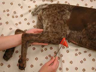
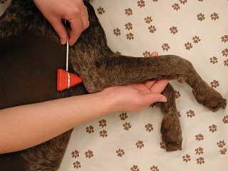
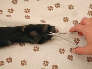
After a complete cranial nerve evaluation has been performed, it is necessary to evaluate the rest of the body and limbs for normal reflexes and responses. Some tests are valuable in evaluating the general function of a large portion of the nervous system, while others help in locating a specific area where the nervous system has been injured. Once a problem has been identified, the help of a local veterinarian should be sought. The veterinarian can help identify the exact location and cause of the problem. The following pictures show how to perform many of these basic tests:
 |
|
|
|
|
If the video
does not play, you must install an MPEG video |
 |
|
 |
|
|
|
|
|
|
If the video
does not play, you must install an MPEG video |
 |
|
Conclusion: The information presented here is not intended to give a complete understanding of a dog’s nervous system or give the individual pet owner enough information to do a complete neurological exam. However, it is intended to provide a pet owner with enough details to recognize a nervous system problem and even begin to locate the injured or diseased area. Additional diagnostics and advice from a local veterinarian are always essential in any problem associated with the nervous system.
* Additional details about different problems with the nervous system can be found on page F548 of this manual.