Introduction:
Several techniques exist which help a medical professional observe the inside of a patient. These techniques are known collectively as diagnostic imaging techniques. One of the most common and routinely used of these techniques is radiography, more commonly referred to as X-rays. A radiograph allows the clinician to look beyond the skin surface to help find clues that can lead to a diagnosis.Description: X-rays are electromagnetic vibrations of very short wavelengths with several unique capabilities. Following are two of these unique features which allow the X-ray to be used for diagnostic imaging:
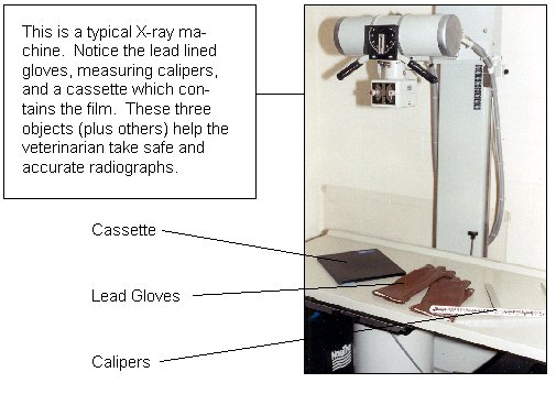
A radiograph machine generates X-rays which are directed through any part of the body. The operator of the machine can control the quantity and speed of the rays being directed through the body part. A radiographic film, protected from light in a flat container known as a cassette, is positioned so that it will "catch" the remaining rays which have penetrated through the body part. The film is then developed through a series of chemical treatments much like everyday photographic film. The final picture depicts dark (radiolucent) areas where a greater portion of the rays were able to penetrate the object and white (radiopaque) areas where the rays penetrated to a lesser extent. Using a quality machine, sensitive film, and the proper technique, great detail can be seen with the use of radiography.
Bone and other dense structures appear white on the film. Gas in the lungs or the digestive tract appears dark. Other organs and tissues have their own unique combinations of dark and light tones, based on their density and the amount of rays that pass through them.
Sometimes a special contrast dye or air can be introduced into various places in the body to help outline certain structures. This type of radiography is known as contrast radiography and is used to examine a body structure in even more detail. This procedure is often performed on the urinary, neurologic, and cardiovascular systems.
Many diseases and problems can be identified by using a radiograph.
Following is a list of some of the situations where a radiograph may be beneficial:Diagnostic Value: Very high. Exact diagnosis can often be identified with radiology. If a diagnosis is not identified, radiographs will often lend suspicion to a diagnosis or identify a problem area requiring additional investigation.
Risk to Patient and Personnel: There is potential risk with repeated exposure to X-rays. X-rays have been proven to be carcinogenic (cancer-causing), and this risk is compounded with repeated exposure. Because of this, special protective clothing must be worn when working with X-rays. Every precaution is taken to avoid unneeded exposure to pet or human. This is accomplished by the use of protective lead clothing, minimizing exposure time and energy to obtain the required radiograph, and by taking the radiograph only when needed. With attention to these few precautions, a radiograph can be very safe and extremely useful. Risks to patients vary because of numerous factors. Sedation may be required for some dogs, and positioning may be uncomfortable or even dangerous for critical patients.
Relative Cost: The relative cost is moderate to high, depending on the extent of the exam, size of the animal, and the number of films taken.
