| A side view of the cow’s reproductive system. |
anatomy | physiology | estrous cycle | reproductive hormones | embryo and fetal development | male reproduction
Introduction:
The difference between a successful dairy operation and one that struggles is often the number of days open the cows experience. The challenge is trying to manage all the possible influences that can affect the reproductive health of a given herd or animal. Each of the following play a role in reproductive health:The information that follows will give the producer a basic background in cattle reproduction and physiology, and then identify the major management issues that need to be considered when evaluating reproductive health.
Female Reproductive System
Terms
:
Male Reproductive System
Terms
:Both Male and Female:
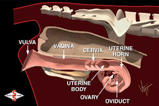
The following text and diagrams will identify the major parts that make up the reproductive system in female cattle. First off, there are two ovaries, two oviducts, two uterine horns, a uterine body, the cervix, the vagina, and the vulva. The bladder lies below the reproductive tract and is connected at the urethral opening located on the floor of the vagina. The rectum is located above the reproductive system.
|
|
The vulva is the external opening to the reproductive system. The vulva has three main functions. It allows the passage of urine out of the body, provides the opening for mating, and serves as part of the birth canal. Included in this structure are the lips and the clitoris. The vulva lips are located at the sides of the opening and appear wrinkled and dry when the cow is not in heat. As the animal approaches estrus, the vulva will usually begin to swell and develop a moist, red appearance. The vagina, about 6 inches in length, extends from the urethral opening to the cervix. During natural mating, semen is deposited in the front (anterior) portion of the vagina. The vagina will also serve as part of the birth canal at the time of calving.
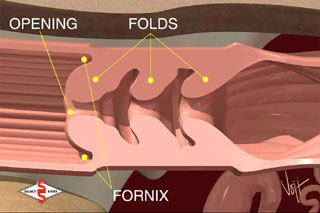
The cervix is a thick-walled organ forming a connection between the vagina and uterus. It is composed of dense connective tissue and muscle that is the primary landmark when inseminating cattle. The opening into the cervix protrudes back into the vagina. This forms a 360° blind ended pocket completely around the cervical opening. This pocket is referred to as the fornix. The interior of the cervix contains 3 or 4 annular rings or folds. This design helps the cervix to protect the uterus from the external environment.
 |
|
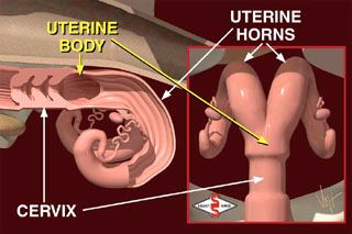
The cervix opens into the uterine body. About an inch long, the body of the uterus serves as a connection between the two uterine horns and the cervix. The uterine body is the site where semen should be deposited during artificial insemination.
From the uterine body on, the reproductive tract separates and all further structures come in pairs. The two uterine horns consist of layers of muscle and a heavy network of blood vessels. The main function of the uterus is to provide a suitable environment for fetal development. When a cow is bred, either naturally or by artificial insemination, the uterine muscles, under the influence of the hormone oxytocin, rhythmically contract to aid in sperm transport to the oviducts.
 |
|
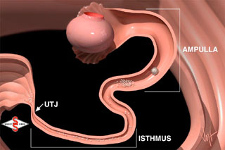
Oviducts, as their name implies, carry the cow's eggs or ova. The oviducts are also commonly referred to as the fallopian tubes. When examined microscopically, the oviduct has several different regions. The lower segment, closest to the uterus, is called the isthmus. The connection between the uterus and the isthmus is called the utero-tubal junction or UTJ. The UTJ functions as a filter of abnormal sperm, and the isthmus acts as a reservoir for healthy sperm.
Research suggests that after gaining access to the isthmus, healthy spermatozoa attach themselves to the walls. During this period of attachment, many physiological changes occur to sperm membranes that are essential to attainment of fertilization potential. These changes are collectively referred to as capacitation and are apparently regulated by this very important attachment to the walls of the isthmus. It takes about 5 to 6 hours after insemination or breeding before a sufficient number of fertile sperm cells can populate the isthmus and complete the capacitation process.
The upper portion of the oviduct, closest to the ovary, is referred to as the ampulla. The interior of the ampulla is more open than the isthmus, allowing for easier passage of ova. It is within this segment of the oviduct that fertilization actually occurs. It is believed that a chemical signal, released at the time of ovulation, stimulates the release of spermatozoa from the walls of the isthmus allowing them to continue their journey to the site of fertilization in the ampulla.
 |
|
Ova are recovered by the large open end of the oviduct that surrounds the ovary. This funnel-like structure called the infundibulum keeps the ova from falling into the body cavity. Hair-like structures on the infundibulum and within the ampulla carry ova and a surrounding mass of cells called the cumulus mass down the oviduct to the site of fertilization.
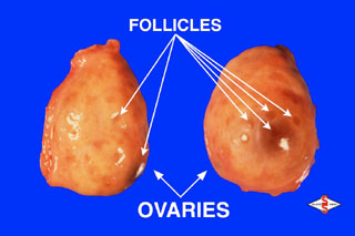
The ovaries are the primary organs in a cow's reproductive tract. They function to produce eggs and to produce hormones (estrogen and progesterone) throughout the different stages of the estrus cycle. Follicles and/or a corpus luteum can often be found on the surface of the ovary. Follicles are fluid filled, blister like structures that contain developing oocytes or eggs. Usually, numerous follicles that vary in size from barely visible to 18-20 mm in diameter can be found on each ovary. The largest follicle present on one of the ovaries is termed the "dominant follicle" and is the most likely candidate for ovulation when the animal comes into heat. Over time, greater than 95% of the other follicles on the ovary regress and die without ovulating and are replaced by new crops of growing follicles.
 |
|
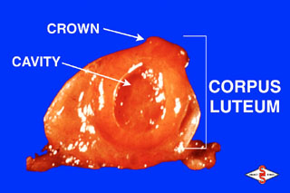
The other structure found on the ovarian surface is the corpus luteum or CL. The CL is the site where ovulation occurred during the previous cycle. Unless there were twin ovulations, only one CL located on one of the two ovaries will be found. The CL will usually have a distinct crown protruding from the ovarian surface that facilitates identification during rectal palpation. The CL may also have a fluid-filled cavity but usually has a much thicker wall than a follicle and thus is a much denser texture. The outside of a CL is usually dark red in appearance, yet a cross section reveals a bright yellow to yellow-orange interior. This is where the term "Corpus Luteum" (Latin for "yellow body") originates.
 |
|
The reproductive stages and cycles in cattle are summarized in the following tables and descriptions:
Table 1 - Female:| Age when puberty reached | Breeding season | Estrous cycle length | Amount of time in estrus | Time when ovulation occurs | Time that corpus luteum (CL) must be present to maintain pregnancy | Gestation length |
| 8-12 months | Non-seasonal (will breed anytime) | 21 days | 12-18 hours | 30 hours after the onset of estrus | Throughout pregnancy | 278-293 days (280 ave.) |
| Age when puberty reached | Breeding season | Type of penis | Volume of semen per ejaculation | Total sperm per ejaculation | Length of time for new sperm to mature (spermatogenesis) |
| 10-12 months | Non-seasonal (will breed anytime) | Fibroelastic with a sigmoid flexure | 2-20 mLs (5 mL ave.) | 2-12 billion (7 billion ave.) | 60-70 days |
Female Reproduction: To fully understand reproduction in female cattle, a basic knowledge of the predominant hormones that are involved is a must. This discussion will identify and discuss these six:
Estrous Cycle: Over a period of time, many changes take place in the reproductive system in response to changing hormone levels. These changes in normal open females repeat approximately every 21 days. This regular repetition is called the estrous cycle. The following will discuss how the estrous cycle works starting with a cow in heat on day zero. On day zero, one ovary has a large follicle approximately 15-20 mm in diameter. This follicle has a mature egg inside ready to be released. The cells lining the follicle are producing the hormone estrogen. Estrogen is transported in the blood stream to all parts of the cow's body, causing other organs to react in a number of ways. It makes the uterus more sensitive to stimulation and aids in the transport of semen at the time of insemination or breeding. Estrogen causes the cervix to secrete mucus that flows and lubricates the vagina. Estrogen is also responsible for all signs of heat including going off feed, allowing other cows to mount, bellowing, holding the ears erect, and a red, swollen vulva.
On Day 1, the follicle ruptures or "ovulates" releasing the egg to the waiting infundibulum. Several hours prior to ovulation, estrogen production declines. As a result, the cow no longer displays the familiar signs of heat. After ovulation, new types of cells (luteal cells) grow in the void on the ovary where the follicle was located.
Over the next 5-6 days, these cells grow to form the corpus luteum (CL). The CL produces another hormone, progesterone. Progesterone from the CL prepares the uterus for pregnancy. Under the influence of progesterone, the uterus produces a nourishing substance for the embryo called uterine milk. At the same time, progesterone causes a thick plug to form in the cervix, preventing access of bacteria or viruses into the uterus. Progesterone also prevents the animal from returning to estrus by blocking the release of follicle stimulating hormone from the pituitary gland in the brain. Follicle stimulating hormone, or FSH, as its name implies, stimulates the growth of follicles. Rapid follicle growth usually results in estrogen production that would bring the animal back into heat and terminate the pregnancy. Thus, progesterone's block to FSH is a very important aspect of maintaining the pregnancy.
Days 16 through 18 of the estrus cycle are referred to as "the period of maternal recognition." During this time, the uterus searches itself for the presence of a growing embryo. If no embryo is detected, the uterus begins to produce another hormone called prostaglandin. Prostaglandin begins to destroy the CL. When the CL is destroyed, no more progesterone is produced and the pituitary gland begins to secrete FSH. This rise in follicle stimulating hormone initiates the selection of a new follicle to grow to a large size, produce estrogen, and bring the animal back into estrus.
A full cycle is now completed. The average total time is about 21 days. The estrus cycle is subdivided into two phases based on the dominate hormone or ovarian structure during each phase. The luteal phase begins when the corpus luteum is formed (about 5 to 6 days after the cow was in heat) and ends when the CL regresses (about day 17 to 19 of the cycle). Progesterone levels are high during this phase of the cycle and estrogen levels are low. The other phase of the cycle is called the follicular phase. It begins when the CL of one cycle is regressed and ends when the new CL of the following cycle is formed. Thus, the follicular phase encompasses the period of time surrounding estrus. During this phase of the cycle, estrogen levels are typically high while progesterone levels are low.
As mentioned earlier, follicles may be present on the ovaries throughout the estrous cycle. Research using ultrasound technology has characterized follicular growth as occurring in "waves." Normally, an animal will have 2 or 3 waves of follicular growth during a 21-day cycle. The beginning of each wave is characterized by the rapid growth of numerous follicles. From this wave or crop of follicles, and by mechanisms still unclear, one follicle is allowed to grow to a much larger size than the others. This large follicle is referred to as the "dominant" follicle because it has the ability to regulate or restrict the growth of all other follicles on the ovary. Dominant follicles only remain dominate for a short period of time (3 to 6 days). This is followed by cell death and regression or ovulation. Consequently, disappearance of the dominate follicle coincides with recruitment of the next crop or wave of follicular growth. From this new wave another dominant follicle will be selected. Although it is typical for follicular growth to occur throughout the estrous cycle, low levels of FSH during the luteal phase, when progesterone levels are high, prevents these follicles from producing high levels of estrogen that would bring the animal back into heat. It is only the dominate follicle present at the time the CL regresses that is permitted to produce enough estrogen to bring the animal back into estrus and to continue on to ovulation.
Summary: In a simplified overview, GnRH is released by the hypothalamus, causing the pituitary to produce FSH. The FSH stimulates the ovary to begin to develop a follicle. As the follicle matures, estrogen is secreted by the follicle and the pituitary is stimulated to produce increased amounts of LH. This increase in LH (LH surge) causes final maturation of the follicle and ovulation of the egg or ovum.
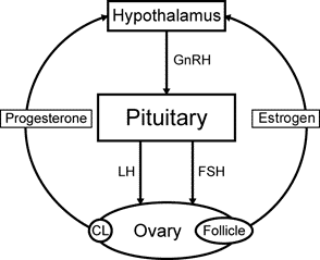
Once a follicle has ovulated, it fills with blood and becomes a corpus hemorrhagicum. The corpus hemorrhagicum becomes a corpus luteum (CL) when the tissues/cells of the ruptured follicle and ovary begin to produce progesterone instead of estrogen. This process of forming a CL is called luteinization.
It is now time for the body to determine if a successful pregnancy has occurred. If the egg or ovum is not fertilized, the female must deteriorate or regress the CL. This is accomplished by the release of prostaglandin F
2a (PGF2a) from the uterus. The PGF2a causes regression (loss) of the CL or luteolysis. In the pregnant female, the fertilized embryo prohibits the release of PGF2a by the uterus. This leaves the CL fully functional and able to produce progesterone necessary for the pregnancy to be maintained. With an understanding of the physiology and related hormones, a discussion of the various hormones used to manipulate a cow’s estrous cycle can now be understood. The following table lists the common drugs used to influence a cow’s estrous cycle: Table 3| Name of Drug | Active Ingredient | Time it should be given in relation to
the cow's estrus cycle. (day 1 = ovulation) |
Desired Result |
| Lutalyse, In-Synch, Prostamate or Estrumate |
Prostaglandin (PGF2a) | Between day 5 and 15 of her estrus cycle. | To remove a CL that is responsive to prostaglandin. It can also be used to clean up an infection in the uterus, to synchronize estrus cycles, and to cause an abortion. |
| Cystorelin, Fertagyl or Factrel | Gonadorelin (GnRH) | Anytime | To treat cystic ovaries and stimulate a cow to cycle. |
Embryo and Fetal Development:
For the first 4-5 days following insemination or breeding, the egg moves in the oviduct toward the uterus. Once the egg gets there, it will be bathed in uterine fluids and continue to grow. While floating free in the uterus, several membranes, including the amnion, the chorion, and the allantois, are produced by the new embryo. Collectively, these membranes are referred to as the placenta. Hopefully, by the time the period of maternal recognition arrives (days 16-18), the fetus and growing placenta will have produced adequate quantities of the chemical signal required to maintain pregnancy. This chemical signal interferes with the action of prostaglandin on the corpus luteum. The CL is thus retained and continues to produce progesterone which is essential to maintenance of the pregnancy.At about 30 days of gestation, the placenta begins to attach to the uterus at several points. The placental sides of these attachment points are called cotyledons, while the uterine side has caruncles. The attachment of cotyledons to caruncles is very similar to velcro. This greatly increases the surface area within the attachment point. Surface area within attachment points is important to allow for exchange of nutrients and waste products between calf and mother. At calving, the muscles in the uterus begin to contract and eventually expel the calf and membranes through a dilated cervix and vagina. Several hormones (progesterone, estrogen, prolactin, relaxin and corticoids) produced by the mother, the fetus, and the placenta, interact to bring about this event.
Male Reproduction: GnRH production by the hypothalamus causes the pituitary to produce FSH and LH. These stimulate the cells of the testicle to produce testosterone, estradiol, and spermatozoa or sperm. Because hormones and other reproductive drugs are seldom, if ever, used in a breeding bull, more attention will be placed on sperm development and factors that influence sperm viability.
|
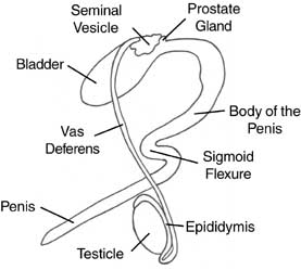
The male reproductive system |
In the tiny tubules (seminiferous tubules) of the testicle, immature sperm are produced. These sperm move to the epididymis, where they fully mature and are stored prior to ejaculation. During ejaculation, the sperm travel through the vas deferens, and into the urethra. In the vas deferens and urethra, additional fluid from the secondary sex glands (seminal vesicles, prostate, bulbourethral glands) is added which helps keep the sperm alive.
Sperm are very sensitive to extremes in heat or cold. Animals that are sick, have a fever, or are fat (causing insulation for the sperm) often have lower numbers of healthy sperm. Severe cold, stress, and pain also cause decreased numbers of normal sperm. Many of these changes to the sperm are not seen until 60 days after the injury/insult occurred (it takes 60 days for sperm to completely mature).
Many of the pictures and much of the text were used with permission from Select Sires, Plain City, OH 43064. Phone # 614-873-4683.