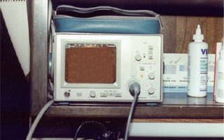
| This is an electrocardiogram (ECG or EKG) machine. This machine is used to determine if a heart condition is causing any abnormalities in the heartís electrical rhythm. It is used to help diagnose specific arrhythmias of the heart. |
ventricular septal defect (vsd) | atrioventricular valve dysplasia | patent ductus arteriosus (pda) | endocardial fibroelastosis | aortic stenosis | pulmonic stenosis | other congenital heart defects | primary (idiopathic) cardiomyopathies | secondary cardiomyopathies | valvular disease | pericardial disease
Introduction:
The heart is an extremely complex organ upon which the life of any animal depends from minute to minute. It is responsible for providing the power to circulate blood throughout the body.Heart problems are relatively common in feline patients seen at veterinary hospitals. Most cats with heart problems are 7 years and older. Some heart diseases, however, are more common in younger cats. Heart disease in cats can be very complicated and must be treated on an individual basis. Therefore, this section will give only a brief overview of some of the more common heart disorders seen in cats. The intent of this material is to give the pet owner a better understanding of the clinical signs, diagnosis, and treatment for many of the problems associated with the feline heart.
Common Terms:
- Right atrioventricular valve (tricuspid valve): Valve which separates the right atrium from the right ventricle.
- Pulmonic valve: Valve which controls the outflow of blood from the right ventricle into the lungs.
- Left atrioventricular valve (mitral valve): Valve which separates the left atrium from the left ventricle.
- Aortic valve: Valve which controls the outflow of blood from the left ventricle into the aorta and the rest of the body.
The "lub-DUB, lub-DUB" sound heard when listening to the heart with a stethoscope is actually the noise made when the valves shut and create turbulence in the blood. The pulmonic and aortic valves close on the "lub" portion of the sound, while the right and left atrioventricular valves close on the "DUB" portion of the sound.
Heart Disease
A large variety of problems can lead to heart disease. Deaths stemming from a heart disorder are usually due to either the development of a fatal arrhythmia or to congestive heart failure and its various complications. All heart problems can be placed into one of two categories: congenital or acquired. A few of the more common heart diseases will be addressed as follows:
Congenital - These are defects present from birth. Congenital cardiac defects are among the most common defects that occur in kittens. This is probably due to the complicated process of cardiac development in the unborn fetus.
- Clinical Signs: Many cats with a VSD do not show significant outward signs of disease, particularly if the defect is small. However, if the defect is larger, the cat may experience signs that include failure to gain weight and thrive, difficulty breathing, and coughing. If Eisenmengerís physiology develops, the cat can have blue (cyanotic) gums.
- Diagnosis: The murmur caused by a VSD is usually found by listening with a stethoscope in the sternum area on the left side of the cat. Most commonly, the condition is detected initially by a veterinarian listening with a stethoscope to an apparently healthy kittenís heart. An ultrasound of the heart (echocardiogram) is necessary to view the shunt. An electrocardiogram (ECG or EKG) is also important to determine if the condition is causing any abnormalities in the heartís electrical rhythm.
- Treatment: Medical therapy initially involves reducing the fluid build-up (edema) caused by the congestive heart failure. This means a salt-restricted diet, rest, and other medications. Surgery is also an option and involves performing a procedure called pulmonary artery banding. This surgery consists of passing band-like material around the pulmonary artery in an attempt to reduce the circumference of the artery. This will increase the pressure in the right side of the heart and reduce the amount of left-to-right shunting blood flow.
* A similar defect can be found in the wall (septum) that separates the left and right atria. This defect is called an atrial septal defect (ASD) and is not as common as VSDs in small animals.
- Clinical Signs: Many cats with atrioventricular valve problems have signs associated with congestive heart failure. These include difficulty breathing, coughing, and a lack of energy.
- Diagnosis: The murmur caused by these problems is usually found by listening with a stethoscope in the sternum area on the left side of the cat. An ultrasound of the heart (echocardiogram) or cardiac catheterization is necessary to specifically diagnose these diseases. An electrocardiogram (ECG or EKG) is also important to determine if the condition is causing any abnormalities in the heartís electrical rhythm.
- Treatment: Medical therapy initially involves reducing the fluid build-up (edema) caused by the congestive heart failure. This consists of a salt-restricted diet, rest, and other medications. Because the surgery to attempt correcting these problems is difficult and rarely done, medical therapy is usually the only treatment.
- Clinical Signs: Most cats with a PDA experience signs that include failure to gain weight and thrive, difficulty breathing, and coughing. Some experience weakness in the hind end and a bounding pulse. Most commonly, the condition is detected initially by a veterinarian listening with a stethoscope to an apparently healthy kittenís heart.
- Diagnosis: The murmur caused by a PDA is unique, notorious for having a continuous or "washing machine" sound. An ultrasound of the heart (echocardiogram) is necessary to view the shunt. An electrocardiogram (ECG or EKG) is also important to determine if the condition is causing any abnormalities in the heartís electrical rhythm.
- Treatment: Surgery is performed in some situations in cats that have no other heart defects and the shunt is still left-to-right. Surgery consists of passing suture (stitch) material around the open ductus and tying it closed. While the procedure may sound simple, it does carry serious risks and must be performed by a skilled surgeon.
- Clinical Signs: Cats with this disease generally begin to show signs when they are 3 weeks to 4 months of age. The first signs that are noticed include difficulty breathing and cyanosis (bluish color to gums) when exerted. At times a murmur is heard when listening with a stethoscope.
- Diagnosis: An ultrasound of the heart (echocardiogram) and X-rays (radiographs) are often helpful in identifying this disease. An electrocardiogram (ECG or EKG) is also important to determine if the condition is causing any abnormalities in the heartís electrical rhythm.
- Treatment: Medical therapy initially involves reducing the fluid build-up caused by congestive heart failure. This prolongs the life of the cat, but affected animals rarely recover.
- Clinical Signs: Abnormal signs are often absent in kittens, but this does not make the disease any less serious. A cat may die suddenly and without warning before its owner is even aware anything is wrong. Fainting spells (syncope) may also occur. These murmurs are often first noticed during a veterinarianís examination of a kitten.
- Diagnosis: The diagnosis of aortic stenosis is made by echocardiology (ultrasound of the heart), through which the thickened tissue ring can be seen and blood pressures inside the heart may be calculated. Chest radiographs and ECG studies may also be helpful.
- Treatment: Treatment includes various medications to help reduce stress on the heart muscle. Some surgical techniques are available, but are difficult to perform, costly, and as a result, are seldom performed. Aortic stenosis is a difficult disease to treat, and the prognosis, even with treatment, is not generally good.
These are disorders which are not present at birth, but develop at some point later in life. These diseases are broken down into four major categories: primary (idiopathic) cardiomyopathies, secondary cardiomyopathies, valvular disease, and pericardial disease. The term "idiopathic" means that there is no known cause for these diseases of the heart.
- Hypertrophic cardiomyopathy is the most common acquired heart disease in cats. This disease results in thickening (hypertrophy) of the left ventricular heart wall. This can obstruct the flow of blood out of the heart, elevate blood pressure in the atria, and cause congestive heart failure. This problem has been associated with high blood pressure and hyperthyroidism in cats.
- Clinical Signs: These signs include collapse, fainting spells (syncope), difficulty breathing, coughing, an increased heart rate, and a lack of energy. In some cats, blood clots may form in the heart and be spread to various parts of the body. A common area for this problem to occur is the arteries which go to the hind legs. Clots that lodge in these arteries cause paralysis of the hind legs.
- Diagnosis: A heart murmur is often heard and X-rays (radiographs) show enlarged atria and a "valentine shaped" heart. An echocardiogram (ECG) is also essential in identifying this disease.
- Treatment: Treatment should focus on reducing the respiratory problems by using drugs that reduce the amount of fluid in and around the lungs. Other drugs should be used to reduce the heart rate and the amount of force the heart expends during its contraction. Aspirin may be used to help prevent blood clot formation. If associated with hyperthyroidism, proper therapy for the overactive thyroid gland may reverse the cardiomyopathy.
- Clinical Signs: Signs of this disease include weight loss, collapse, fainting spells (syncope), difficulty breathing, coughing, increased heart rate, fluid accumulation (edema), and hind limb paralysis from blood clot formation.
- Diagnosis: A heart murmur is often heard with a stethoscope and X-rays (radiographs) show an enlarged heart and left atrium. An echocardiogram is the best technique for identifying this disease.
- Treatment: Treatment should focus on reducing the respiratory signs by using drugs that reduce the amount of fluid in and around the lungs. Other drugs should be used to maintain a regular heart rhythm and prevent ventricular tachycardia (ventricles beating too fast). Aspirin can be used to help prevent blood clot formation.
- Endocrine (hormone related) - hyperthyroidism, excessive growth hormone secretion
- Genetic - atrioventricular myopathy
- Infiltrative (invasive) - neoplasia
- Inflammatory - viral, fungal, bacterial, or protozoal infections
- Lack of blood flow (ischemia) - blood clots (thromboembolism)
- Nutritional - taurine and L-carnitine deficiency
- Pressure or volume overload on the heart - high blood pressure (hypertension) and valve problems
- Toxic - rattlesnake venom and doxorubicin (chemotherapeutic drug)
*The diagnosis and treatment for each of the above causes of secondary cardiomyopathy will be different depending on the cause. In general, the symptoms affecting the cat will need to be treated and the cause of the condition will need to be eliminated if possible.
- Infective Endocarditis - This is an infection of the heart muscle, valves, or supporting structures of the heart. Bacteria are usually responsible for the infection, although fungal infections can also occur. The bacteria must first enter the bloodstream and be carried to the heart. Because the bloodstream carries bacteria to all parts of the body, organs other than the heart (particularly the kidneys and the spleen) may also be affected. The source of bacteria entering the bloodstream differs from individual to individual; however, the most common sources are the mouth, skin, digestive tract, and urinary tract. Cats with bad teeth, urinary tract infections, ear and skin infections, or that will eat anything, including garbage, are more at risk.
The heart valves and supporting structures seem to be the more common sites of infection within the heart, possibly due to their constant exposure to turbulent blood. Any of the four heart valves may be affected. Valves on the left side of the heart (aortic and mitral valves) are affected more often than the valves on the right side of the heart.
- Clinical Signs: Heart murmurs (especially a new heart murmur never previously heard), fever, and sometimes lameness are found in cats with infective endocarditis. Other clinical signs will vary and depend upon where in the heart the infection has occurred and what other organ systems are involved.
- Diagnosis: Blood cultures are an important part of making the diagnosis of infective endocarditis. This diagnostic test requires that blood samples be taken and sent to a diagnostic laboratory for culture. The culture results may take up to 3 weeks to be finalized; therefore, treatment is usually started before a final test result can be obtained. If bacteria are found circulating in the bloodstream, and recent heart problems are present, a positive (definitive) diagnosis of infective endocarditis can be made. If the actual bacterial colony is large enough, it may be viewed with echocardiography (ultrasound of the heart). Radiographs (X-rays) of the heart, CBC, blood (serum) chemistry panel, and ECG are other recommended tests to aid in diagnosis. See Section D for additional information on the above tests.
- Treatment: Therapy for infective endocarditis focuses on antibiotics and heart stabilization. Based on their general success in helping patients with this condition, antibiotics are chosen in the beginning before blood culture results can be finalized. After obtaining blood culture results, a veterinarian will select antibiotics based on the culture and sensitivity data (please see Culture and Sensitivity page D135). Treatment for damage done to the heart itself is based on severity, structures affected, and need.
- Prevention: Maintaining a healthy pet is critical in preventing this disease. While nothing will guarantee 100% prevention, providing basic pet hygiene is perhaps the most important part an owner can take in avoiding this ailment. Brushing a catís teeth regularly and providing annual dental cleaning under the care of a veterinarian are central in pet hygiene. Keeping a close watch for skin and ear infections, anal gland impactions, urine abnormalities, and strictly monitoring diet are also important components of pet hygiene. Each of these areas is given special attention in other sections of this manual.
- Pericardial effusion - This is fluid that accumulates around the heart in the "heart sac" (pericardium). This fluid can accumulate because of low protein levels, heart failure, certain cancers, bacterial infections, viral infections, or fungal infections. In cats this fluid can also accumulate because of feline infectious peritonitis and toxoplasmosis infections. If the fluid accumulates rapidly, a situation called "cardiac tamponade" can result. Cardiac tamponade, a life threatening problem, is a sudden compression of the heart from rapid fluid accumulation in the pericardium.
- Clinical signs: Common problems include difficulty breathing, weakness, lack of energy, collapse, and distension of the abdomen. In cases of cardiac tamponade, the cat will have a weak, rapid pulse, distended veins, and diminished heart sounds when listened to with a stethoscope.
- Diagnosis: Rhythm abnormalities are common and the heart can appear round in shape and enlarged when observed using a radiograph. The most accurate way of diagnosing this problem is by echocardiography.
- Treatment: In severe cases the fluid must be removed by performing a procedure called a pericardiocentesis. This is where a sterile catheter is used to cleanly enter the pericardium and remove the fluid. The cause for the fluid build-up must be identified and then treated. Depending on the cause, treatment may include antibiotics, antifungals, fluid reducing drugs, anti-cancer agents, plus others.

|
|