F610
Orthopedic Problems
problems found in the front limb |
problems found in the hind limb |
problems found in the spine | problems found in the skull
| arthritis | degenerative joint disease
| anti-inflammatory drugs |
infectious arthritis |
immune-based arthritis |
growing bone diseases |
neoplasia | fractures |
classification of fractures |
managing fractured bones |
infections | osteomyelitis
| septic arthritis
Introduction: Orthopedics is the branch of medicine and surgery that
focuses on the health of the bones and associated tissues. There are many types
of orthopedic problems that affect cats. Many of these incidents are minor
problems that can resolve on their own with rest and time. Minor sprains,
twists, or muscle bruises account for a large percentage of these common
problems. Persistent lameness, any lameness associated with trauma, and severe
lameness problems should always receive the attention of a veterinarian.
Recovery from an orthopedic problem may take weeks to months.
Locating the problem area and potential cause: Limping or lameness is one
of the more common problems encountered by cat owners. Often, the challenge
comes when trying to determine the location and then the cause for the lameness.
At first, it is important to consider the following questions. These are also
questions that a local veterinarian may ask when evaluating the animal:
- What is the activity level of the cat?
- Have there been any prior surgeries or medical problems?
- What is the animalís age, sex, breed, size, diet, and life-style? (For
example, an outdoor cat that frequently gets into cat fights is much more
likely to sustain a bite wound that becomes infected and leads to lameness
than a cat which is kept strictly indoors.)
- Is the cat currently on any medications?
- Has the condition come on suddenly or slowly?
- Does the problem seem to be associated with an injury?
- Does the problem get worse after exercise?
A thorough physical and orthopedic examination should then be performed. The
goal of an examination is to identify the location of the lameness. The
following are some basic steps that are often used when performing an orthopedic
exam:
- The cat should be observed while standing. Signs of muscle shrinking
(atrophy), confirmation abnormalities, swelling, and pain can often be found.
- Next, the patient is observed from the front, side, and rear while
walking and possibly jumping up onto a chair or table. When the lame limb
contacts the ground, the stride of the affected limb is usually shortened as
compared to the opposite, normal limb. When the sound limb hits the ground,
the animal will often spend more time with that limb in contact with the
ground during the walking motion. Other problems that are often noticed
include ataxia (lack of coordination), paralysis, paresis, and short, choppy
gaits. If more than one limb seems to have a problem, a more central condition
may be at fault. It is important to remember that a perfectly normal gait
requires the use of almost the entire nervous system and many of the muscles
and bones of the body. Injury, damage, or tumors that affect the nervous
system, muscles, or bones can cause problems in one, two, or all four limbs.
- Once the problem limb(s) is identified, a thorough examination of the
limb(s) is necessary to further localize the problem. The examination must be
systematic, starting at one end of the limb and proceeding up or down, feeling
every bone and every joint in the limb. Each area of the limb, including
muscles, bones and tendons, is felt (palpated) as gently as possible until the
painful area is identified. All this should be done while the animal is fully
awake without any sedation. Sedation may mask the animalís response to the
testing. Each joint is moved through the entire range of motion by flexing,
extending, and rotating. Each joint should also be felt for evidence of
swelling, pain, heat, lack of motion, instability, crepitus (popping), and
laxity.
Abnormal laxity (slackness) may indicate
ligamentous injury or damage to the joint capsule. Some fractures are often
very obvious, while others may only be identified by excessive movement of the
area, pain, swelling, and lack of use of the limb. A hot, swollen joint often
is the result of an infection causing inflammation. If the injury has caused
the animal to avoid using the limb(s) for a period of time, the muscles may
shrink (atrophy). Injury to muscles is often identified by tenderness,
swelling, pain, and heat.
- After a specific area of injury has been identified, radiographs
(x-rays) are often used to help determine the extent of the damage and
determine the appropriate treatment.
Note: Basic bone and muscle anatomy of the cat can be found on page
A34
of this manual. Pictures of actual radiographs showing different orthopedic
problems can be found throughout the following information. The cost involved in
treating an orthopedic problem may be substantial. However, the outcome is often
good to excellent with proper treatment and patience.
Many of the most common orthopedic problems in cats are included in the
following information. Each disease or problem is categorized in this
information based on where it is located on the body. The front limb will be
covered first, followed by the hind limb. Diseases that affect more than one
area of the skeleton will then be covered separately.
Problems Found in the Front Limb:
Major orthopedic problems that commonly affect the front limb include
fractures, ligament and tendon injuries, growing bone diseases, dislocations,
infections, and cancers.
- Shoulder: The shoulder joint is the connection between the
scapula (shoulder blade) and the humerus (upper leg). Dislocations and
fractures in this area do occur but are unusual in cats because the shoulder
is well-protected by surrounding muscles and other tissues. Arthritis can also occur in the shoulder joint.
- Shoulder joint dislocation can occur as a result of trauma in
cats. X-rays are usually needed to tell the difference between a shoulder
dislocation and a fracture. Treatment always requires professional help.
Sometimes the dislocation may be successfully treated by moving the bones
back into place and having the cat wear a special bandage for
2 weeks. Often, however, surgery is a necessary part of the treatment.
Upper leg (humerus): The humerus is the long bone extending from
the shoulder to the elbow. Fractures of the humerus are uncommon in cats. The
radial nerve is one of the major nerves in the front limb and travels directly
across the surface of the humerus. This nerve can be damaged when the humerus
is fractured. Fractures of the humerus are easily diagnosed with x-rays and
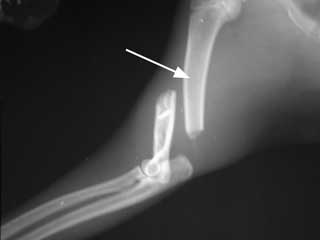
always require surgery to repair (see figure #1).
 |
|
|
| Figure 1: Fractured Humerus -
This is a fracture in the lower third of the humerus. The white arrow
indicates the upper portion of the humerus that connects with the
shoulder. This type of fracture would need to be repaired surgically. |
|
Elbow: The elbow joint is a "hinge" joint connecting the humerus
of the upper front limb to the radius and ulna of the lower front limb. The
elbow is relatively unprotected and can be easily damaged and fractured.
Dislocations of the elbow joint can also occur but seem to be less common than
fractures. Arthritis (see below) of the elbow joint can occur,
especially following trauma or injury to the joint.
- Elbow joint dislocation is uncommon but can occur as a result
of severe trauma. The joint is quite stable, and severe trauma is more
likely to result in a fractured bone than in a dislocation of the elbow.
X-rays are needed to tell the difference between fractures and dislocations
of this joint; occasionally, both a fracture and dislocated elbow will be
seen together following a severe blow to a front leg.
Treatment of dislocations requires setting the bones in their proper
positions while the cat is under anesthesia. If the procedure can be done
within a few hours after the trauma, the setting of the bones can often be
performed without surgery. The more time that has passed since the
dislocation occurred, the more difficult it becomes to place the bones back
in their normal positions. Surgery is necessary to set the bones if they
cannot be replaced in their normal positions with anesthesia alone.
Placement of a splint or bandage after setting the bones in place may be
helpful to achieve rapid healing. Arthritis is common later in life in any
joint that has been dislocated.
Lower leg (radius/ulna): The radius and ulna are paired bones
that connect the elbow to the carpus or wrist joint. The radius is the major
weight-bearing bone of the two. Fractures of the radius and/or ulna are fairly
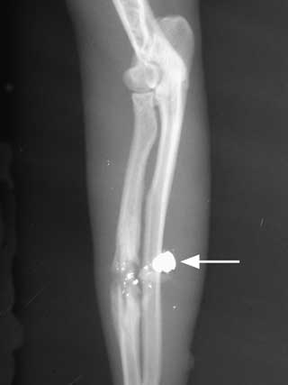
common in cats (see figure #2). When fractures of the lower front limb occur,
both bones are usually broken together. However, it is not uncommon to have a
fractured radius and an intact ulna following trauma to a front limb. X-rays
are usually not needed to know whether a fracture of the lower front limb is
present but are important to determine the nature of the fracture. Many fractures require surgery, while others can be properly treated with
a cast or splint to stabilize the fractured bone(s).
 |
|
|
| Figure 2: A fractured radius
caused by a bullet (white arrow). |
|
Carpus (wrist): The carpus is a very complex structure. There are
seven small bones in two rows that connect the lower front limb to the paw and
make up the carpal joints. There is a separate joint connecting each row of
bones, making a total of three joints in the carpus. Injuries to the carpal
joints and bones are not uncommon in cats. Most of these are soft tissue
injuries, involving the ligaments and joint capsule only. X-rays are always
needed following injury to this area to examine the small bones of the carpus
and to determine if any fractures exist. Fractures can occur in any of the
bones of the carpus, although they are infrequent. Dislocations of the carpus
can occur along any of the joints. If there is severe damage to ligaments and
other structures that support the carpus, fusion of the joint (a surgical
procedure called arthrodesis) may be necessary.
- Dislocation of the carpus can occur at any of the three joints
as a result of traumatic injury to the front limb. Damage to the ligaments
and other tissues that support the carpus is usually difficult to repair and
does not heal very well. Special x-ray pictures can help diagnose a
dislocation at one or more of the three joints in the carpus. A dislocation
of any or all of these is generally termed "hyperextension." When
weight-bearing, a hyperextended carpus allows the paw to drop closer to the
ground than normal. The condition is usually quite painful. Minor cases of
hyperextension of the carpus may be successfully treated with a splint or
bandage and strict rest. However, most hyperextension situations require
more aggressive treatment. Fusion of the joint, a surgical procedure known
as "arthrodesis," is necessary in many cases to restore function.
Paw: The feline front paw is made up of five digits (toes), four
of which are weight-bearing. The dewclaw, which corresponds to the human
thumb, sits high up on the inside surface of the front paw and does not bear
weight. There are five bones called metacarpals that extend from the carpus to
each of the five digits. Three bones make up each of the four weight-bearing
digits. These bones are called the first, second, and third phalanges. The
dewclaw has only two phalanges. The nail bed is a delicate tissue arising from
the upper surface of the last phalanx of each digit, and from this tissue each
nail grows. A thick, rubbery pad grows on the underside of each paw, and a
smaller pad grows on the underside of each of the digits. Lameness that
results from pain in the paw is common. Infections of the skin between the
digits and around the pads, abrasions of the pads themselves, and a variety of
cuts and scrapes all may result in pain and lameness. Fractures of the
metacarpals and the digits are also quite common. Finally, cancers of the paw
are rare, but can occur in the nail beds of cats. These nail bed tumors have
usually spread to the nail beds from some other location. Life-threatening
lung and skin cancers have been reported to spread to multiple nail beds in
some cats.
Problems Found in the Hind Limb:
Major orthopedic problem categories that commonly affect the hind limb
include fractures, ligament and tendon injuries, growing bone diseases,
dislocations, infections, and cancers.
- Pelvis: The pelvis is a bony structure responsible for
transferring the weight of the hind end to both hind legs. The pelvis is
divided into different areas. Weight is first transferred from the lower spine
to bones on the right and left sides. These bones are called the ilia
(singular = ilium). The ilia transfer weight to the hip joints. Connecting the
right and left sides of the pelvis are two other bone sets known as the pubis
and the ischium. The ilia, pubis, and ischium together make up the pelvis, a
box-like structure. The acetabulum is the socket-like portion of the ilium
that connects to the femur.
By far, the most common problem encountered involving the pelvis is trauma
and injury. Some estimations report that nearly 25% of all fractures involve
the pelvis. Other problems that may involve the pelvis include dislocations,
infections of the bone or surrounding tissues, and cancers.
- Pelvic fractures are extremely common with trauma of nearly any
type. Because of the rigid box-like shape of the pelvis, fractures are
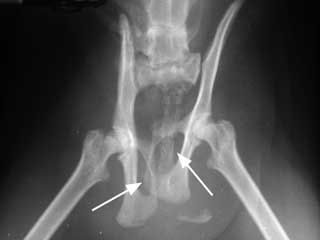
always multiple and usually occur in sets of three (see figure #3). Pelvic
fractures range from minor to extremely devastating. Because the right and
left ilia are the main weight-supporting portions of the pelvis, fractures
of these bones are usually very serious. Fractures of the socket-like
acetabulum that houses the ball of the hip joint are also extremely serious
and usually result in arthritis of the hip later in life, regardless of how
well they heal. Fractures of the pubic bone are often minor and are allowed
to heal without intervention. Fractures of the ischium vary in severity but
are quite often allowed to heal on their own because they usually do not
affect the animalís ability to bear weight.
Diagnosis of pelvic fractures is made with x-rays. Treatment depends
completely on the situation. Many minor to moderate pelvic fractures can
heal very well with strict cage rest and anti-inflammatory pain killers
alone. More serious types of fractures, such as those that affect the hip
joint or cause changes in the natural shape of the pelvis, should be
treated with surgery. Surgery for pelvic fracture repair is extremely
difficult and is always expensive. Anti-inflammatory pain killers and
laxatives to help reduce pain associated with the passage of bowel
movements are important aspects of the treatment plan for many pelvic
fracture victims.
 |
|
|
| Figure 3: Fractured Pelvis -
Notice how the femurs and obturator foramina (white arrows) do not
line up. When the pelvis is fractured, it usually breaks in at least
three places. |
|
- Dislocations of the pelvis can occur with trauma. The only
place in the pelvis where a dislocation may occur is at the attachment of
the ilium to the spine. This is known as the "sacro-iliac joint." A strong
ligament that attaches the ilium to the spine may be torn with severe
trauma.
Like pelvic fractures, pelvic dislocations are diagnosed with x-rays and
treated according to severity and situation. Surgery is often the preferred
type of treatment for pelvic dislocations.
Hip: The hip joint consists of the "ball-and-socket" structure
connecting the pelvis to the hind limb. The femur is the first long bone in
the hind leg that connects the pelvis to the knee or stifle joint. The femur
is the longest bone in the body. As previously indicated, the medical term for
the socket is the acetabulum. The medical term for the ball is merely the
"head" of the femur. A variety of conditions can affect the hip joint.
Fractures and dislocations are common.
- Dislocation of the hip joint is a common result of injury to
the hind end in cats. Blunt force trauma causes the ball to pop out of the
socket, tearing the ligaments and capsule that help keep the joint in place.
Diagnosis of this condition can often be made through examination by a
veterinarian. Confirmation of the dislocation is needed with an x-ray (see
figure #4). The x-ray provides important information for proper treatment
because it will also show the veterinarian what type of dislocation has
occurred. Treatment consists of getting the hip joint back together and
keeping it there. This can be more challenging than it sounds. If action is
taken quickly, the head of the femur may be put back into place by working
the ball back into the socket. This is performed by a veterinarian while the
cat is under anesthesia. Because more inflammation in the injured area
actually "locks" the bones in their abnormal positions, this procedure
becomes increasingly more difficult with the passage of time. If the
dislocation cannot be repaired in this manner, surgery becomes necessary. A
variety of methods are available, depending on the situation.
 |
|
|
| Figure 4: Dislocated Hip- The
white arrow identifies the head of the femur that is dislocated out of
the hip socket (acetabulum). Compare this side to the opposite hip
that is not dislocated. |
|
A special bandage called an Ehmer sling is usually placed on the leg
regardless of the type of treatment used. This sling helps to keep the hip
in place while preventing the cat from putting any weight on the leg for up
to 2 weeks.
- Hip dysplasia is far less a problem in cats than it is in dogs,
but is reported in the Siamese breed. It appears to be passed in families
and can result in crippling lameness. Animals with hip dysplasia are born
with normal hip joints, but changes occur during development and aging.
Gradual loosening of the joint with swelling, pain, and damage to joint
tissues occurs.
Diagnosis of the condition must be
made with x-rays and can be treated medically with anti-inflammatory
medications. Surgical treatment is the most effective and rewarding
treatment available. Total hip replacement can be done in cats as it is in
dogs; however, because the condition is seen in cats on a less frequent
basis, the surgery is not routinely performed. A less expensive alternative
to the total hip replacement surgery is called the femoral head and neck
osteotomy (FHO), in which the head and neck of the femur ("ball" of the
ball-and-socket) is completely removed and the upper portion of the femur
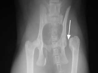
allowed to form another "socket" joint with scar tissues (see figure #5).
The surgery is remarkably effective with lightweight animals, and cats tend
to do extremely well following this procedure.
 |
|
|
| Figure 5: Femoral head and neck
osteotomy (FHO) - The white arrow shows the location where the head
and neck of the femur used to be located. An FHO surgery has been
performed. |
|
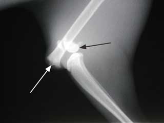
Upper leg (femur): The femur is the long bone extending from the
hip to the knee (stifle). Fractures of the femur are common. Fractures of the
femur are easily diagnosed with x-rays and always require surgery to repair
(see figures #6-7). Repair is often very difficult and can be very expensive.
There are many different ways of repairing a fractured femur, so an orthopedic
specialist should be consulted.
 |
|
|
| Figure 6: Fractured Femur -
This is a fracture of the femur just above the bottom or distal end of
the bone. The femur is indicated by the white arrow and the black
arrow identifies the distal end of the femur or the condyles. To
adequately fix this type of fracture, orthopedic surgery is required. |
|
 |
|
|
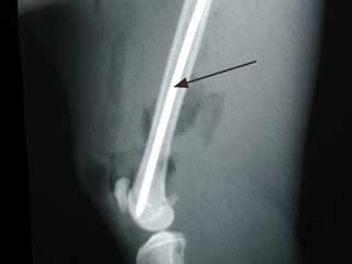
| Figure 7: The black arrow
identifies a bone pin placed to repair the fracture shown in figure #6
above. |
|
Stifle (knee): The stifle joint is another hinge joint like the
elbow but vastly different in many ways. The lower portion of the femur ends
at the stifle joint. On the front side of the lower end of the femur sits the
patella (knee-cap), a small bone that moves up and down as the stifle bends.
Small cushions of cartilage sit in between the lower end of the femur and the
upper end of the tibia. These cushions of cartilage are called menisci (the
lateral meniscus on the outside and the medial meniscus on the inside). Two
ligaments run down the sides of the stifle joint, and two ligaments criss-cross
inside the stifle joint. All these parts are important in the function of the
stifle joint, and all can be injured and lead to pain and lameness.
Injury to the stifle joint is uncommon in cats. When injuries do occur to
this joint, they may involve fractures, ligament injuries, or dislocation.
Probably the most common injury sustained to the stifle joint is the tearing
of one of the inner criss-cross ligaments. The ligament commonly injured is
called the cranial cruciate ligament and is identical to the anterior cruciate
ligament in the human knee joint. Improper movement of the patella or knee-cap
(called patellar luxation) may also occur. Arthritis of the stifle is seen
frequently in older patients, usually as a result of some previous injury or
problem in the joint. Bone cancers, while rare in cats, may affect the stifle
joint area.
- Cranial cruciate ligament injuries occur uncommonly in cats.
When the cranial cruciate ligament (CCL) tears, this allows the tibia to
slide forward away from the femur too much when the animal bears weight on
the leg. This excessive movement stretches the other tissues in the stifle
area, resulting in pain. The sliding movement also damages the cartilage
cushions (menisci) that sit between the two bones. Most cats with a torn CCL
are very reluctant to bear any weight on the leg. The lameness can occur
suddenly, although many cats will have been exhibiting an on-off mild
lameness for weeks to months before. Tearing can happen in an otherwise very
healthy stifle joint with a sudden and severe blunt force to the area. More
commonly, however, the ligament will gradually weaken and deteriorate as the
animal ages. Aged, weak ligaments will often tear easily with minor force,
such as slipping or jumping. In many of these cases, an owner will not know
of any trauma received by the pet. It is important to be aware that these
ligaments age and can become weak in both stifle joints over time, and when
the CCL tears on one side, there is a chance that the other will also tear
in time. This likelihood is probably made even greater with the extra weight
borne by the "good" leg when one ligament tears. Overweight cats may be more
likely to suffer CCL tears for the same reason.
Diagnosis of CCL tears is made during
a detailed exam by a veterinarian. The forward movement of the tibia is
detected and helps determine the diagnosis. Some cats may need to be sedated
for a thorough examination if they are too tense or in too much pain. X-rays
can also be helpful in showing inflammation in the stifle joint but cannot
show the actual torn ligament. Torn cranial cruciate ligaments should always
be repaired with surgery. If not repaired surgically, the joint does
stabilize itself the best it can with thickening of tissues. The cat will
improve gradually up to a limited point after many months of lameness.
However, arthritis will always result and is often severe. Thus, if left
alone and not repaired with surgery, the cat will usually appear to improve
for awhile but will end up getting permanently worse in the end. Once the
arthritis sets in, it is irreversible. Prognosis with surgery is usually
good.
- Patellar luxation (dislocation of the knee-cap) is another
uncommon condition of the stifle joint of cats. This condition is seen most
commonly in the Devon Rex and domestic shorthair breeds. The patella usually
dislocates towards the inside of the leg (medially) and is generally
apparent by the time the animal is 4-6 months old. Both stifle joints are
usually affected.
Trauma to the stifle can also cause dislocation of the patella in an
otherwise healthy cat. Patellar instability is graded based on its severity.
Grade 1 is the most mild form, while Grade 4 is the most severe. The more
mild cases will often gradually progress to become severe later on. Some
cats will adapt to the patella popping in and out of place as they walk.
Some cats may hide this problem so well that the owner may not notice any
lameness until the problem is very severe. Arthritis of the stifle is
usually a consequence of patellar dislocation.
Diagnosis of this problem is made primarily by physical examination. A
veterinarian can detect the abnormal movement of the patella and determine
the severity of the problem. X-rays are helpful with the diagnosis,
especially in showing if any arthritis is present.
Treatment depends on the situation. Mild cases with no lameness present
are monitored for worsening of the condition. Severe cases require surgery.
There is a large gray zone in the middle of these two extremes. Treatment
for cases falling in this gray zone, with only occasional lameness and/or
moderate instability, must be judged individually by the owner and the
veterinarian. Surgery can be extremely helpful for one particular individual
but may not work at all in another.
- Stifle dislocation is a very serious problem where numerous
ligaments and other tissues have been severely damaged. It requires a great
deal of force to cause dislocation of the stifle joint. Dislocation of the
stifle joint usually includes tearing and severe damage to both cruciate
ligaments and at least one of the menisci. Diagnosis of this problem is made
by careful examination by a veterinarian while the animal is under
anesthesia. X-rays are very helpful as well, showing damage to parts of the
joint that cannot be examined from the outside. Treatment is best
accomplished by carefully reconstructing the joint. Such a surgery may be
lengthy and very expensive. If reconstructive surgery is successful and
proper care is taken afterwards for healing, the outcome is often
surprisingly good. Mild arthritis and/or stiffness of the joint may result.
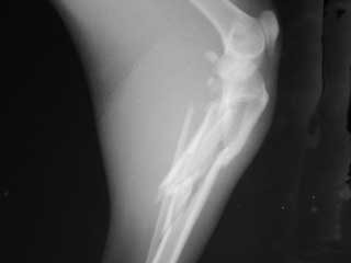
Lower leg (tibia/fibula): The tibia and fibula are paired bones
that connect the stifle to the tarsus or ankle joint. The tibia is the major
weight-bearing bone of the two. Fractures of the tibia and/or fibula are
extremely common in cats. When fractures of the lower hind limb occur, both
bones are usually broken together (see figure #8). However, it is not uncommon
to have a fractured tibia and an intact fibula (or vice versa) following a
hind limb trauma.
X-rays are usually not needed to know
whether a fracture of the lower hind limb is present but are important to
determine the nature of the fracture (see below). Treatment of certain
cases requires surgery, while some fractures may be properly treated with a
cast or splint to stabilize the fractured bone(s).
 |
|
|
| Figure 8: Fractured Tibia and
Fibula - This is a comminuted fracture of the tibia and fibula. |
|
Tarsus (ankle): The tarsus or hock joint is a very complex
structure. There are seven small bones in two rows that connect the lower hind
limb to the paw and make up the tarsal joints. There is a separate joint
connecting each row of bones. The calcaneus (heel bone) is the largest of
these bones and projects back, toward the catís tail. The Achilles tendon
(common calcaneal tendon) attaches to this bone and travels up to the muscles
on the back of the catís lower limb. The Achilles tendon is rather exposed and
can be injured easily. Injuries to the hock joints and bones are common in
cats. Most of these are soft tissue injuries, involving the ligaments and
joint capsule only. X-rays are always needed following injury to this area to
help evaluate the small bones of the hock and to determine if any fractures
exist. Fractures can also occur in any of the bones of the tarsus. Such
fractures are uncommon. Dislocations of the tarsus can occur along any of the
joints. If there is severe damage to ligaments and other structures that
support the tarsus, fusion of the joint (a surgical procedure called
arthrodesis) may be necessary.
- Dislocation of the tarsus can occur at any of the joints as
a result of traumatic injury to the hind limb. Injuries to ligaments in the
joint and sometimes fractures of the small bones are associated with hock
dislocation. Diagnosis is made through careful examination by a veterinarian
and x-rays. Replacement of damaged ligaments, repair of fractured bones, or
fusion of the joint, also known as arthrodesis, may be necessary to restore
function.
- Severing of the Achilles tendon is possible as a result of
blunt trauma or sharp injuries to the hind limb. If the tendon has actually
torn, surgery will be necessary to repair it. Some cases respond to
splinting or casting the leg at its full stretched length, which helps the
tendon to pull tight again as it heals.
Paw: The feline hind paw is usually made up of four digits, all
of which are weight-bearing. The first digit, the dewclaw, which corresponds
to the human thumb, sits high up on the inside surface of the hind paw and
does not bear weight. The hind dewclaws are missing at birth in cats. The
anatomy and problems associated with the hind paw are identical to the front
paw.
Problems Found in the Spine:
The feline spine is divided into five sections. First, the cervical or neck
portion consists of seven vertebrae (bones that make up the spinal column). The
first cervical bone that connects to the base of the skull is called the atlas,
and the second cervical bone is called the axis. These two bones are distinct
from all other vertebrae in their shape. These bones, along with the other
vertebrae of the spine, provide protection for the delicate spinal cord that
runs through their center, while at the same time allow for some limited
movement in the neck and back. The second section of the feline spine is called
the thoracic spine and consists of 13 vertebrae. The 13 ribs connect on either
side of the chest to these vertebrae. The third section is called the lumbar
spine and consists of seven vertebrae that correspond to the "lower back" in
people. Three fused vertebrae make up the unique section of the spine called the
sacrum found in the pelvic part of the spine in cats. The final section of the
spine is made up of a varying number of vertebrae that become smaller and
smaller as the tail tapers. These are called the coccygeal or tail portion of
the spine. Vertebrae have projections called processes that extend dorsally (up)
and laterally (sides). The processes are termed dorsal spinal processes and
lateral spinal processes. They vary in length and shape depending upon where in
the spinal column they are located. The dorsal spinal processes are the small
ridges that can be felt running the length of the back. These are very useful in
helping to count the vertebrae in the spine during examination.
Spinal column injuries can be devastating to an animal because any damage to
the delicate spinal cord running through the center of the spine can result in
permanent paralysis of portions of the body. Intervertebral disk disease, where
the cushion-like disks in between the bones of the spine become deformed, is not
commonly seen in cats. Spinal injuries and diseases are more commonly associated
with the branch of medicine entitled neurology rather than with orthopedics. The
most common type of abnormality associated with the spine in cats is probably
the kinked tail, a birth defect which is rather common among some breeds and in
some geographical areas. This defect can occur anywhere along the length of the
catís tail, but is more commonly found toward the very end. Corrective surgery
of the problem is usually not effective.
Problems Found in the Skull:
The cat skull is extremely complex. It is made up of several bone plates that
fuse together as the cat matures, as well as the two paired mandibles or
jawbones with their hinges in the back of the lower skull. The central portion
of the skull houses the brain and brainstem that are critical to the function of
the entire body. Located in front of the brain and on the sides of the face near
the nose are open compartments known as sinuses. Hollow openings at the front of
the skull house the eyes. The paired mandibles from which the lower teeth grow
are connected to each other at the very front part of the skull by a strong
ligament and are hinged at the lower back portion of the skull by a joint known
as the temporomandibular joint (TMJ). The right and left maxillary bones make up
the sides of a catís nose and are connected to each other on the bottom by the
palatine bone, hard palate, or roof of the mouth. The upper teeth grow from the
lower portion of the maxillary bones as it attaches to the hard palate.
Injuries to the skull occur relatively infrequently and are usually
associated with blunt trauma, such as an automobile accident. Fractures of any
bone in the skull can occur, and depending on what organs or soft tissues are
involved, these fractures can be very serious. Head trauma with severe brain or
brainstem injury is usually fatal. Most other injuries to the mouth, nose, and
face are treatable, although permanent disfigurement can result. Because the
many different injuries that can occur to the head, each type of injury must be
managed on an individual basis and under the care of a veterinarian.
Arthritis:
Arthritis simply means inflammation of a joint. Arthritis can be broken down
into several different categories based on the type, location, and cause of
joint inflammation. Joints can be freely mobile, partially mobile, or immobile.
While inflammation can occur in any joint, the freely mobile joints and, to a
lesser extent, the partially mobile joints are those most likely to create
problems for the body when they do become inflamed.
The disk joints in between vertebrae in the spine are the most common
partially mobile joints where problems develop in the cat. Intervertebral disk
disease, where the cushion-like disks in between the bones of the spine become
deformed and cause back pain and sometimes paralysis, is not common but does
occur. Freely mobile joints include all the joints of the limbs such as the
shoulders, elbows, hips, stifles (knees), and all the joints in the paws
("knuckles"). While the specific makeup of each of these joints differs, each
contains the same basic structures.
A thin layer of cartilage overlying the bone in each joint provides for
frictionless motion. A thick, tough wall called the joint capsule attaches to
the bone on all sides and protects the delicate cartilage from damage. The joint
capsule also holds in the sticky, clear lubricating fluid that is found inside
the joint. This fluid is called synovial fluid and also helps with joint
lubrication and frictionless movement.
The three broad categories of arthritis seen in cats include degenerative
arthritis, infectious arthritis, and immune-based arthritis. Cats are fortunate
in that all three categories are relatively uncommon.
- Degenerative joint disease (DJD) is the medical term for the
degenerative type of arthritis that occurs in joints. Specific types of DJD
have already been discussed, such as hip dysplasia. The main
features of DJD are the wearing away of the cartilage and the formation of new
bone at the joint margins. Degeneration of a joint can occur as a result of a
long list of causes. Genetics, nutrition, previous injury to a joint or nearby
structures, infection, and age are some of the more common reasons for
developing this type of arthritis. DJD often gradually gets worse with time
and may become extremely severe and even crippling.
Clinical signs of this degenerative arthritis are generally limited to
signs of pain: limping, stiffness, reluctance to exercise, protectiveness and
even hissing or biting when painful areas are touched. In severe cases, the
patient may stumble or fall, slip easily on stairs or slippery surfaces, or
may become unable to walk at all if more than one limb is affected.
Because of the decrease in activity often associated with arthritis, cats
may become overweight. This creates what is known as a "vicious cycle," where
the additional weight puts extra strain on the joints, which makes the
arthritis worse. This causes the cat to exercise or move even less, leading
back to more weight gain, and so on.
Diagnosis is based upon physical examination and x-ray films. Treatment for
degenerative joint disease is complex. One approach available for some types
of arthritis is surgery. Femoral head and neck ostectomy (FHO) for hip
dysplasia is a very good example of surgical treatment for
arthritis of the hip joint. Other types of surgery may be helpful, such as
joint fusion or joint reconstruction. Decisions on whether surgery is an
option for treating joints already affected with DJD are best made with the
help of an orthopedic specialist.
Medical therapy for DJD is currently a
very complex topic. Many cats with degenerative arthritis will benefit from
medical therapy to some degree, and in many cases the improvement is dramatic.
Much of this improvement is due to general pain relief. Another benefit of
medical therapy is slowing down the progression of disease. There are two main
groups of medications that are used to treat degenerative arthritis. The first
group consists of anti-inflammatory drugs that help reduce pain and restore
function. The second group consists of nutritional supplements containing
natural hormones and other products thought to protect the joint tissues from
damage and even change and improve the makeup of the joint tissues and fluid.
Anti-inflammatory drugs have been used
over the years in the treatment of joint pain and arthritis in cats.
Anti-inflammatory drugs come in two types: steroids and non-steroids.
Non-steroidal anti-inflammatory drugs (known by the acronym NSAIDs) include
aspirin, flunixin meglumine, phenylbutazone, ketoprofen, and meloxicam. NSAIDs
are becoming widely recognized as an important part of the treatment of
degenerative arthritis. As cats become arthritic, the pain of movement
naturally leads to decreased exercise and activity. Weight gain then becomes a
common problem in less mobile pets. Extra weight increases stress on the
arthritic joints, leading to increased inflammation and pain. The additional
pain then causes the cat to become even less mobile, and the "vicious cycle"
mentioned previously continues. Perhaps the greatest benefit of NSAID use in
cats with arthritis is the breaking or hindering of this cycle. Use of
anti-inflammatory pain killers can keep an arthritic cat moving and
exercising, thus maintaining a healthy weight and slowing down the progression
of joint disease.
Many different types of NSAIDs have been
listed previously. This is not an indication that all are recommended or
healthy for use in cats. In fact, NSAIDs tend to have a rather narrow margin
of safety, which means that it is quite easy to create problems with them.
Cats are very sensitive to many of these drugs and they should be used with
caution and only under the direction of a veterinarian. Acetaminophen
(Tylenol), for example, can cause death in cats. NSAIDS which have been used
safely in cats include aspirin, phenylbutazone, meloxicam, flunixin, and
ketoprofen. Side effects of NSAID use include vomiting, listlessness, loose
stools, and hives.
Steroids are often used for their powerful anti-inflammatory effects in
cats, particularly because they tend to be safer than most NSAIDs. Cats are
also much less likely to experience side effects such as weight gain from
steroid use than other species.
- Infectious arthritis is the condition where an infection leads to
inflammation of a joint. There are several different types of organisms that
can infect the joints. Examples of bacterial infections, protozoal infections,
and fungal infections of joints will be discussed.
- Septic arthritis is the condition where a joint becomes
infected with bacteria, usually from a penetrating injury or bite wound.
This particular type of arthritis is discussed
below.
- Lyme Disease: Lyme disease is caused by a bacterium known as
Borrelia burgorferi and is transmitted by ticks of the Ixodes family.
Cats have a much greater resistance to Lyme disease infection than do dogs.
Rarely, lameness due to Lyme disease infection may occur in cats. Diagnosis
is generally made with serology. Treatment is effective with antibiotics,
such as amoxicillin or tetracycline. No vaccine currently exists for
prevention of Lyme disease in cats. Please see page F498 for additional
details on Lyme disease.
- Ehrlichiosis: Feline ehrlichiosis is caused by several species
of small bacteria in the rickettsial family. While the disease in uncommon,
it is seen occasionally in the western and midwestern United States. The
disease can affect all body systems, with arthritis in multiple joints being
a common clinical sign. Fever, loss of body condition, swollen lymph nodes,
pneumonia, and blood disorders may also result from feline ehrlichiosis.
Diagnosis is based on bloodwork (serology). Treatment with antibiotics is
generally very effective. Doxycycline, an antibiotic similar to
tetracycline, is the antibiotic of choice for treatment of feline
ehrlichiosis.
- Toxoplasmosis: Toxoplasmosis in cats is caused by a protozoal
parasite called Toxoplasma gondii. Generally, young or stressed cats
are the most likely to show clinical signs of disease; healthy adult cats
may become infected, but do not often show any signs of disease. Infection
occurs through ingestion of the parasite in hunted prey such as birds or
mice. A form of infective arthritis may occur along with many other clinical
signs in affected cats. Diagnosis of toxoplasmosis may be made with
bloodwork. Treatment of choice includes antibiotics (clindamycin) and
sometimes supportive care. Please see page F836 for additional details.
- Fungal arthritis: Fungal joint infections most often occur
secondary to fungal bone infections (see below under "Infections"). A
variety of fungal types may infect the joints of cats. Diagnosis is best
made with special testing of the joint fluid. A lengthy treatment with
antifungal drugs is often necessary.
- Immune-based arthritis is the third category of arthritis in
cats. The term "immune-based" indicates that these types of arthritis involve
the animalís own immune system destroying the joint tissues. Immune-based
arthritis is typically a disease of multiple joints.
- Chronic Feline Progressive Arthritis: This is a disease in cats
that is not caused by any type of infection, but may be closely associated
with feline leukemia virus (FeLV) infection. About 60% of cats with chronic
feline progressive arthritis are also infected with FeLV. The disease occurs
in 2 different and distinct forms: Rheumatoid-like progressive arthritis and
fibrous ankylosing progressive arthritis. Affected animals may have other
clinical signs such as fever, decreased appetite, and weight loss. Each form
will be discussed below.
- Rheumatoid-like progressive arthritis received its name because of
its similarity to rheumatoid arthritis in dogs and people. It occurs more
commonly in older cats and causes unstable, deformed, and painful joints.
Similar to rheumatoid arthritis, radiographs show erosion of the bones in
the affected joint where cartilage attaches. Rheumatoid factor, a
measurable substance in the bloodstream of dogs and people with rheumatoid
arthritis, is not present in cats, however.
- Fibrous ankylosing progressive arthritis occurs almost exclusively
in young male cats. Joints are swollen, painful, and stiff. New bone
growth tends to fuse joints and severely restrict their normal range of
motion (ankylosis). X-ray films show new bone extending around the joints
with loss of bone structure in general inside the joints themselves.
Diagnosis of both forms of this arthritis is based primarily on history,
physical examination, radiographs of the joint, and testing of the joint
fluid. Steroid therapy with prednisolone and other immune-suppressing drugs
such as azathioprine and cyclophosphamide are the standard treatment.
Prognosis is usually guarded to poor because the disease progressively
worsens to the point of crippling lameness.
- Systemic lupus erythematosus (SLE) is an uncommon disease in
cats that affects multiple systems in the body at one time. Disorders of the
nervous system, respiratory system, kidneys, skin, muscle tissue, blood, and
joints are commonly seen with SLE. Multiple joints typically become swollen
and painful. Affected animals may be listless, have a decreased appetite,
and may have a high temperature. If the kidneys begin to fail, an increased
thirst and need for urination may develop. Skin problems are also common.
Because SLE tends to involve so many systems, diagnosis of this disease
is challenging. Tests that may be helpful in the diagnosis of SLE include
x-ray films, joint fluid tests, and bloodwork. Testing for antibodies the
immune system creates to attack other tissues of the body, also known as "autoantibodies,"
is used in other species to diagnose SLE, but is very unreliable in cats.
Treatment is generally focused on suppressing the immune system with
steroids and other drugs. The expected outcome is not usually good and
becomes very grim if the kidneys begin to fail.
- Idiopathic polyarthritis is a "catch-all" category where all
other unknown causes of multiple joint pain and swelling are placed. Many of
the arthritis cases placed into this category appear to result from illness
elsewhere in the body or because of reactions to drugs and/or vaccines.
Infections in various places including the lungs, tonsils, urinary tract,
skin, and eyes have been linked with idiopathic polyarthritis. Disease of
the digestive tract including inflammation of the stomach, intestines, and
colon may be another cause of idiopathic polyarthritis. Some types of cancer
may also lead to inflammation of the joints. If arthritis of several joints
occurs due to any of these underlying conditions, the treatment must focus
on the specific condition. If an underlying cause can be treated and
resolved, the arthritis will usually go away. Some of these cases respond
well to treatment with steroid therapy.
Growing Bone Diseases:
Diseases of the developing bones are rare in cats. These diseases can be
present at birth or become apparent as the kittenís skeleton develops. Treatment
for these conditions is difficult and often not possible. Examples of some
unusual disorders which may be encountered in kittens include the following:
- Ectrodactyly: This condition, also known as "split-hand
deformity," results from the failure of proper growth or fusion of the bones
in the front paw, leading to a deep cleft in the paw itself.
- Osteogenesis imperfecta: This is a disorder of bone formation at
the microscopic level. It frequently results in deformed limbs, fragile bones,
and multiple fractures.
- Multiple cartilaginous exostoses (MCE): This disease causes
aggressive swelling and growth of bones near the growth plates. Some of the
swellings may become cancerous with time. This disease is listed with growing
bone deformities in general; however, it usually develops in cats after the
skeleton is already mature. There appears to be a strong correlation between
MCE in cats and infection with feline leukemia virus (FeLV). Some references
may categorize this disease with feline bone cancers.
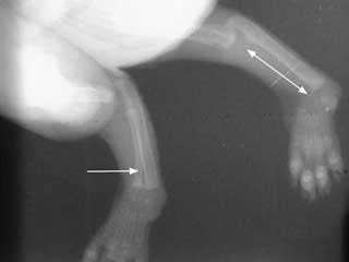
- Radial agenesis: This is a deformity in which the radius never
develops at all in the forelimb and the ulna is thicker and more curved than
normal (see figure #9).
 |
|
|
| Figure 9: Radial Agenesis - The
white arrow with only one point shows the relatively normal radius on
one limb. The double headed arrow shows the place where the normal
radius should have been on the opposite limb. |
|
Neoplasia:
Neoplasia or cancer of the bones is rare in cats. The most common bone cancer
diagnosed in cats is osteosarcoma.
- Osteosarcoma is the most common bone cancer in cats and arises
from bone cells. This type of bone cancer is very aggressive in the bone
itself, but does not spread to other parts of the body as commonly as the same
type of cancer does in dogs or humans. The hind limbs are affected with
osteosarcoma about twice as often as the front limbs. The average age for cats
to develop osteosarcoma is around 10 years old; however, it has been reported
in cats ranging from 1-18 years.
The most obvious clinical signs of osteosarcoma include pain and swelling
in the affected area. When the bone cancer occurs in one of the limbs,
lameness is often very obvious early on. Depending on where the cancer occurs,
swelling of the affected area may become apparent to an owner. Another
clinical sign may occur where the cancer, after weakening the bone from which
it grows, allows the bone to break through the weakened area. This is known as
a pathologic fracture (a fracture of a bone resulting from some underlying
cause). When such a fracture occurs, sudden lameness, inability to bear
weight, pain, and swelling at the fractured area result.
Diagnosis of osteosarcoma begins with x-rays of the affected bone. Trained
professionals can come to a very high degree of suspicion for osteosarcoma
just by looking at a good quality x-ray. For a complete diagnosis to be made,
a biopsy of the cancerous tissue must be sent to a laboratory for
histopathology. A bone biopsy is a tricky procedure with the undesirable side
effect of further weakening the diseased bone. Treatment for osteosarcoma is
so aggressive and drastic, however, that an accurate diagnosis is very
important. The benefits of knowing the diagnosis outweigh the risks of
obtaining a bone biopsy in most cases. Once the diagnosis of osteosarcoma is
made, treatment should be started as soon as possible. Treatment of
osteosarcoma begins with aggressive removal of the affected bone. Amputation
of the entire affected limb is the standard approach for osteosarcoma of the
long bones of the legs. If there is no evidence of spread of the cancer to
other body systems, amputation alone without chemotherapy or other treatment
is often very effective and even considered to cure the cancer in some cases.
- Osteochondroma is a primary bone cancer that arises from
cartilage cells within the bone. A form of osteochondroma known as
osteochondromatosis can occur in which the cancerous growths may arise from
many areas of the skeleton at once. At first the bone growths are usually
benign, but with a fairly good chance of becoming malignant and spreading
cancer throughout the body. There is a possibility that osteochondromatosis is
correlated with feline leukemia virus (FeLV) infections. Diagnosis is made
with physical examination, x-rays, and biopsy/histopathology of the growths.
Treatment is difficult, especially for osteochondromatosis where the cat may
also suffer from FeLV infection.
- Fibrosarcoma is a cancer arising from connective tissue found
throughout the body, including bone tissue. It occurs only rarely in bone and
may be difficult to treat. Diagnosis is made by bone biopsy. Little is known
about how fibrosarcoma of the bone behaves in cats. Aggressive surgery is
considered the best treatment.
- Hemangiosarcoma is a cancer of blood vessel cells that has been
reported as a primary bone cancer in cats. Little is known about how it
behaves, and aggressive surgery is recommended. It has been reported to
metastasize to other sites of the body.
Secondary bone cancers: Tumors that spread from other areas of the body
to bone are called secondary bone cancers. Spreading can occur through the blood
supply to bone from distant places in the body or from tumors that grow right
next to the bone and invade it. Most commonly, secondary cancers occur in bone
from nearby tumors that invade and attack all tissues nearby. Sometimes the
source of the cancer is not known until a biopsy is obtained of the affected
bone and a veterinary specialist finds, for example, cancerous thyroid gland
cells within the bone. Treatment of spreading secondary bone cancer depends on
the nature of the cancer. Treatment of the original tumor is usually of more
importance than treatment of the secondary bone cancer. Surgery, chemotherapy,
or radiation therapy may be feasible treatment options.
Fractures:
Broken bones are the most common orthopedic problem encountered in cats.
Falls, gunshot injuries, and automobile accidents are all common causes of
broken bones in pets. This section will address the basics of dealing with
fractured bones in a practical sense for cat owners.
- Classification of fractures:
- Closed or simple fracture: This is a fracture of a bone where
the skin is not broken and no exposure to the outside environment has
occurred. The opposite is an open or compound fracture.
- Open or compound fracture: This is a fracture where exposure
with the outside environment has occurred. Examples include gunshot injuries
to bones (see figure #2) and fractures where the sharp bone
fragments have cut through the skin.
- Transverse fracture: This is a fracture where the bone is
broken into two pieces and the fracture crosses the bone in a straight,
side-to-side line.
- Oblique fracture: This is a fracture where the bone is broken
into two pieces and the fracture crosses the bone in a diagonal line.
- Spiral fracture: This is a fracture where the bone is twisted
apart.
- Comminuted fracture: This is a fracture where the bone is
broken into multiple pieces.
- Greenstick fracture: This is a fracture where the bone is
broken on one side, with the other side bent but not fractured. This type of
fracture is most commonly seen in young kittens.
- Pathologic fracture: This is a fracture due to a weakened bone
structure from any disease process (i.e. bone cancer, infection, or
osteoporosis).
- Stress fracture: This fracture results from repeated force to a
bone.
- Segmented or double fracture: This is a bone that is broken in
two or more different places.
- Managing fractured bones: The first part of treatment of a broken
bone is often done by the catís owner. Since most fractures are associated
with some kind of trauma, checking the vital functions of the injured pet
should always be done first. Alertness, breathing status, and mucous membrane
color/capillary refill time should be observed quickly and thoroughly. The
petís responsiveness and state of awareness can help assess whether the brain
and central nervous system are functioning on a basic level. Difficulty
breathing may result from internal bleeding, broken ribs, or other injuries.
Pale gum color with a slow capillary refill time occur with severe blood loss
and shock. These are potential life-threatening injuries that must take
precedence over any broken bones. Rapid transport to a veterinary hospital is
very important for trauma patients. Specific home treatment for a broken bone
can be done if there is time and if the patient is cooperative. Handling an
injured pet should always be done with cautionĖ even the most trustworthy pet
may bite or injure its owner when it is hurt. If a wound is present in
association with a broken bone, a clean bandage may be placed on the injury.
Direct pressure should be used to control any bleeding.
Once the patient is brought into the
veterinary hospital, the veterinary staff takes over, repeating the steps
listed above. Beginning with the vital signs, the patient is examined
thoroughly. Life-threatening injuries are given attention first, followed by
fractured bones. A patient must be stabilized before a broken bone is given
treatment; this stabilization process may require hours to days. A bandage is
usually placed on a fracture to help control pain and prevent further injury
until proper treatment can begin. Once the patient is stable, plans for
treating broken bones may be made.
Specific treatment for a broken bone
depends greatly upon the situation. The location and type of fracture,
availability of specialist help, nature of the patient, and cost are all
factors that influence how a broken bone will be treated. Some veterinarians
specialize in orthopedic surgery and should become involved in the more
complex bone injuries. Because these injuries usually require a great deal of
time, personnel, and training to properly treat, the cost of treating broken
bones is generally high.
There are various methods of fixing
broken bones. The broken bone must be placed into the proper position and held
there with some type of supporting device. Splints and casts are rigid
supports that encircle the broken limb and can be used for a variety of
fractures, but have limitations on how much they can do. These types of outer
support can be used with or without internal support for the broken bone,
depending upon the situation.
Other types of support for broken bones may require extensive surgery.
Metal pins inserted into the bone, wires that encircle the fracture and
tighten against the outside of the bone, and metal plates that cross a
fracture and are screwed into the bone are all common types of internal
support that are used. External skeletal fixation is another type of support
that is becoming more common in veterinary medicine. This type of support is
both internal and external because several metal pins are drilled at a right
angles to the bone with the pin sticking out of one and sometimes both sides
of the leg. These pins are then connected to each other with another metal rod
or bone cement, creating a support on the outside of the body. The type of
support used to treat any given fracture depends upon many factors and is best
left to the discretion of the veterinarian.
Infections:
Infections of bones and joints can occur in cats. Infection of bone tissue is
called "osteomyelitis" and infection of a joint is called "septic arthritis."
- Osteomyelitis is the medical term for bone infections. Bone
infections may involve bacteria, fungi, or viruses. Bacteria are responsible
for most bone infections seen in cats. Any cat may be affected, and any part
of the skeleton may become infected. Bone is typically well-protected and
resistant to infection. Open fractures, where the broken ends of the bone
penetrate the skin, are perhaps the most common way the infection reaches the
bone. Infections may also enter bone tissue following surgery, from infected
wounds near the bone, or from the blood supply. Chronic long-term bone
infections can result from open fractures and bone surgery. If a fragment of
bone dies and is left in the fracture area, new bone may form around it and
trap it inside the healed bone. The immune system cannot fight infection
inside of dead tissue since it is cut off from the bodyís blood supply. Thus
the trapped piece of dead bone provides an excellent home for bacteria and
leads to long-term infection. Diagnosis of osteomyelitis is usually made with
a good history, physical examination, and x-rays (see figure #10). Treatment
of osteomyelitis depends upon the situation. Antibiotics and surgery are the
primary means of treating bacterial bone infections. Surgery is needed in most
cases of long-term osteomyelitis to remove pieces of dead and severely scarred
tissue. Antibiotics are provided for several weeks to months.
Fungal bone infections are very uncommon in the cat, but have been
reported. Feline histoplasmosis (Histoplasma capsulatum), a deep fungal
infection, is the most common one found in the United States. Other deep
fungal infections which affect cats include Cryptococcus neoformans and
Blastomyces dermatidis.
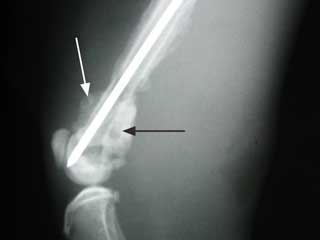 |
|
|
| Figure 10: Osteomyelitis- The
white and black arrows point to areas of osteomyelitis surrounding the
end of this femur. There is also a pin in the femur that was used to
repair a fracture. |
|
- Septic arthritis is the term used for an infected joint. Injuries
to joints are the most common reason for infection to reach these
well-protected areas. Other sources of potential joint infections include
injections directly into a joint, surgery, and access through the blood
supply. The affected cat is usually in pain, reluctant to bear weight, and may
have a fever. The joint may be hot and swollen. Diagnosis of a septic joint is
achieved by history, physical examination, x-rays, and by testing a sample of
the joint fluid. The joint fluid analysis is the most helpful of the tests in
showing the actual infection. Culture and sensitivity (see page
D135) may be
performed on the fluid, often providing very helpful information for
treatment. Septic arthritis is a difficult problem to treat. There is usually
extensive and permanent damage done to the delicate tissues of the joint,
which results in permanent pain and arthritis even after the infection is
gone. Surgery to relieve swelling and pressure, flushing the joint with
sterilized fluid, and removing any clumps of infected material is usually the
best approach. Sometimes, the joint must be left open and carefully bandaged
to allow it to drain. Administration of antibiotics for at least 4 weeks is
very important regardless of the surgical approach. The outcome is very
questionable, and permanent damage resulting in arthritis of the joint is not
uncommon.
Summary: Orthopedic problems are a common occurrence in veterinary
medicine. The key to successfully treating these problems is early detection and
proper treatment. The petís owner plays an essential part in identifying
problems early and then seeking the proper medical attention. In general, if
lameness in a pet lasts longer than 24 hours, the lameness is getting worse, or
there is an obvious injury, veterinary advice should be sought.


