|
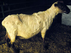
Typical abscesses associated with the superficial form of CLA. |
Introduction/Causative Agent:
Caseous lymphadenitis (CLA), or cheesy gland, is a chronic, contagious disease of sheep and goats caused by the bacterium Corynebacterium pseudotuberculosis. It results in abscesses of the lymph nodes and, less frequently, the internal organs. Once an animal becomes infected, it can easily spread the disease to other sheep or goats through direct contact or by contaminating the environment. Because the organism can survive for long periods of time in the soil and surrounding environment, eradication of CLA can be a lengthy and frustrating process. However, eradication should be considered because of the economic impact and human health risks of the disease. The lesions associated with CLA can be found in animals less than 6 months old, but may occur and recur throughout the lifetime of an animal. As the animal ages, the chances of developing the disease increase. This is most likely due to repeated exposure to the organism.Economic Importance: Losses from CLA are difficult to quantify and are due to a large number of factors. The visceral form (abscesses of internal organs) is the cause of significant economic loss in the sheep and goat industries due to condemnation of carcasses. One source ranks CLA as the third most significant cause of condemnation at slaughter. The superficial form (abscesses of lymph nodes and other areas just under the skin) is the cause of abscess scars that decrease pelt value. Other causes for loss include decreased weight gain, wool growth, milk production, and reproductive efficiency. In addition, affected animals are unthrifty, frequently culled prematurely, and occasionally die. Increased somatic cell counts in dairy animals and decreased value of breeding stock have also contributed to economic losses.
Clinical Signs: Two forms of the disease have been described. The superficial form involves abscessed and enlarged lymph nodes that are located closest to the skin surface. Most commonly, the lymph nodes around the head (intermandibular, parotid, retropharyngeal, and cervical) and lymph nodes near the origin of the limbs (prescapular, prefemoral, and supramammary) are affected. The second or visceral form of the disease involves abscesses of the internal organs and lymph nodes and is often associated with long term debilitation. The organs most likely to be affected are the lungs and associated lymph nodes, kidney, liver, and mesenteric lymph nodes. Both forms are found in sheep and goats; the superficial form is more common in goats, and the visceral form is more common in sheep.
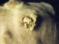
The involved lymph nodes are distended by a thick and often dry discharge (purulent exudate) that is a white, yellow, or greenish color. If external lymph nodes are affected, there is a well-defined, non-painful swelling that grows larger over time. If left untreated, the skin covering the node will become thin and the abscess will eventually rupture to the outside, releasing exudate and bacteria into the environment.
|
|
|
|
Disease Transmission: In both sheep and goats, direct contact with the bacteria results in infection. Such contact can occur when the infectious material of a draining abscess from one animal touches another animal, allowing the bacteria to enter through the skin or mucous membranes. Because the bacteria are capable of surviving for extended periods of time (months to years) in the environment, surfaces can harbor organisms and continuously infect animals as they come in contact with them. It is believed that feed bunks become easily contaminated, particularly in goat herds, resulting in the high occurrence of abscesses around the head and neck in this species. In sheep, the bacteria are spread most commonly at shearing through contaminated equipment. Contaminated sheep dips have also been implicated. Crowding during shearing is another mode of transmission.
Numerous routes of infection have been studied experimentally. Researchers have shown that the organism is capable of entering the lungs by inhalation and can spread to internal organs by injection into the bloodstream. Studies also show that it can cross the membranes of the digestive tract and vagina, and that a break in the skin is not needed for an animal to become infected.
Once the organism enters the body, it travels to local lymph nodes where it forms an abscess. The bacteria release an exotoxin (a toxic phospholipase D or PLD) that causes an intense inflammatory reaction and enables the bacteria to move into the lymph nodes. The healing process causes the development of a thick wall surrounding the bacteria. The bacteria continue to multiply inside the abscessed lymph node. With time, many of the abscesses rupture, causing the bacteria to contaminate the environment and other animals.
Diagnosis:
The superficial form of CLA is usually diagnosed based upon the presence of abscessed lymph nodes and culture of C. pseudotuberculosis from abscess contents. Enlargement of one or more superficial lymph nodes with discharge is suggestive of the disease, but should not be the only criteria for diagnosing this disease.A necropsy may reveal internal abscesses that can also be cultured for confirmation. If CLA is suspected, a gram stain to reveal gram positive, club-shaped rods may aid in determining if an animal should be isolated while awaiting culture results. There are also blood (serologic) tests available that can detect animals infected with early (no abscess formation) or internal forms of CLA. There is a wide variation in the accuracy and cost of these tests. False positives will occur in animals that have been vaccinated against CLA.
Other Causes of Abscesses and Disease:
Staphylococcus spp., Streptococcus spp., and Actinomyces pyogenes also cause abscesses. It is important to confirm that the cause is truly Corynebacterium pseudotuberculosis before initiating stringent control or eradication measures. When the internal form is present, a chronic wasting disease may be all that is apparent. In such cases, it is important to consider the possibility of other problems such as abnormal or poor teeth, Johnes disease, pasteurella pneumonia, ovine progressive pneumonia, scrapie, internal parasites, neoplasia, or inadequate nutrition.Treatment:
Currently, there is no effective antibiotic therapy for CLA due to the way the animal encapsulates the bacteria within the lymph node and isolates it from the rest of its body. Treatment of this disease should always be accompanied by control methods to prevent the spread of the bacteria to other animals. In all circumstances, any affected animals should be immediately isolated. The abscess can be lanced and flushed daily with a strong povidone iodine solution, or the entire lymph node can be removed surgically, thereby eliminating environmental contamination. Some veterinarians successfully treat non-food (meat or milk) producing animals by injecting formalin into the abscess. In most cases, culling the animal is the safest and only option.Vaccination:
There are different types of vaccines now available for immunization against C. pseudotuberculosis. One contains a toxoid of C. pseudotuberculosis alone. Another combines C. pseudotuberculosis, Clostridium perfringens type D, and Clostridium tetani. Although there is little research data currently available to confirm their effectiveness, reports indicate that vaccines help prevent and lessen the effects of the disease. Vaccination alone, however, should not be utilized as the only protective measure against CLA. In most cases, the first vaccination can be given at 3-4 months of age in animals where the mother has previously been vaccinated. In lambs/kids from mothers that have not been previously vaccinated, a first dose should be given at 1-3 weeks of age. Many products should be boostered again 4 weeks later and repeated annually, thereafter, about 6-8 weeks before the lambing/kidding season. Some vaccines may cause the formation of a sterile granuloma at the injection site. This granuloma may persist for many years. Some vaccines can cause significant problems in goats already infected with CLA and should be used with caution.Control and Eradication: An effective control program for this disease requires a great deal of dedication, patience, and daily observation. Eliminating the organism from an infected herd will primarily involve eliminating it from the environment. The following goals are essential when attempting to control CLA and prevent additional infections:
Zoonotic Potential: Corynebacterium pseudotuberculosis infections in humans are rare but can occur, particularly where infected sheep and goats are skinned by hand. The most common scenario involves puncturing of the human skin with infected knives. The disease has also been spread to humans consuming raw milk from infected sheep and goats. Precautions should always be taken to prevent human skin contact with the discharge (purulent exudate) from an abscess or contaminated objects, and milk should always be pasteurized prior to consumption.